

Introduction
External or internal tooth discoloration represents a significant aesthetic concern, particularly when it affects the anterior teeth. Since color is a critical factor in aesthetic dentistry, especially in the anterior region, resolving discoloration after endodontic treatment has become a notable clinical challenge.
One approach to the aesthetic rehabilitation of discolored teeth is the use of ceramic restorations. These are minimally invasive procedures. However, when attempting to restore discolored teeth using veneers or crowns, there is a substantial risk that the dark underlying tooth structure will show through the relatively thin and translucent ceramic. Consequently, several strategies have been employed to address the issue of masking discoloration. These include the use of more opaque ceramics and luting cements, or increasing the depth of tooth preparation. Each of these methods presents its own challenges. Opaque ceramics and cements often result in a flat, non-vital appearance. Deeper tooth preparation to achieve greater ceramic thickness can expose dentin, which provides weaker adhesion. Therefore, it is recommended that preparation design remain within the enamel layer as much as possible to ensure optimal bonding.
Tooth shade assessment is one of the most critical components in restorative dentistry. Discoloration of one or two anterior teeth can have a significant aesthetic impact due to mismatched coloration with adjacent teeth. The eLAB system and other shade quantification platforms may pave the way toward establishing a new standard of best practices in objective shade communication in dentistry. The precise selection from a broad range of customized ceramic powders typically relies on image analysis, individualized shade mapping, and, above all, personal preference and clinical experience. This complexity introduces a significant degree of uncertainty regarding the predictability of shade matching in clinical reality. The present article demonstrates the restoration of anterior teeth with varied shades using the eLAB prime system.
Clinical Case
A 43-year-old female patient, I., presented to the clinic seeking aesthetic rehabilitation of the anterior teeth (Figure 1). Tooth 21 had undergone endodontic treatment more than six years prior. At the time of the initial examination, the tooth was restored with a temporary composite crown. Tooth 11 exhibited a darker shade compared to the maxillary lateral incisors. Following a discussion of the treatment plan, the patient opted for the restoration of teeth 13 to 23 using ceramic restorations.
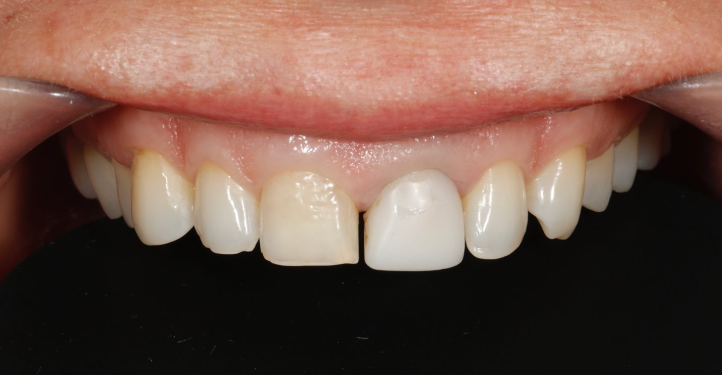 Figure 1. A – anterior teeth with retractors. Tooth 21 previously underwent endodontic treatment and restored with a temporary crown.

B – patient’s smile.
At the second visit, a diagnostic wax-up was performed and discussed with the patient. She expressed a desire to retain the provisional restorations for further evaluation of the shape and contour (Figure 2).
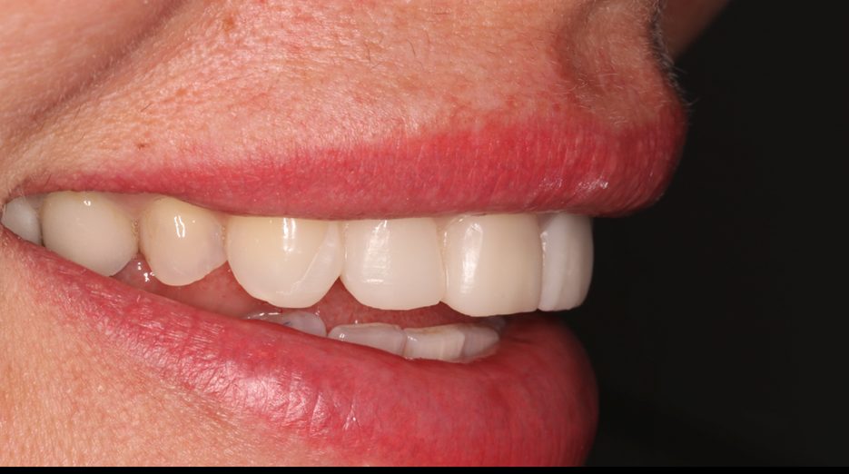
Figure 2: Temporary restorations were placed for shape matching.
At the third visit, tooth preparation for ceramic restorations was performed under magnification using a dental microscope. Initial tooth mapping was carried out through the provisional restorations, followed by the main preparation phase, guided by silicone keys. Tooth 21 was prepared for a full-coverage crown (Figure 3). Subsequently, shade evaluation was conducted for both the unprepared and prepared teeth designated for ceramic restorations (Figure 4). After impression taking, provisional chairside restorations were fabricated using Luxatemp A1 (DMG, Germany).

Figure 3: Stages of tooth preparation. A – tooth marking through temporary restorations

B – view at the impressions stage.

C – Check the with a silicone index.
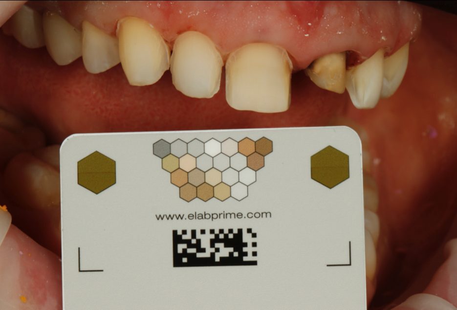
Figure 4. A – photo with the eLAB prime gray card.
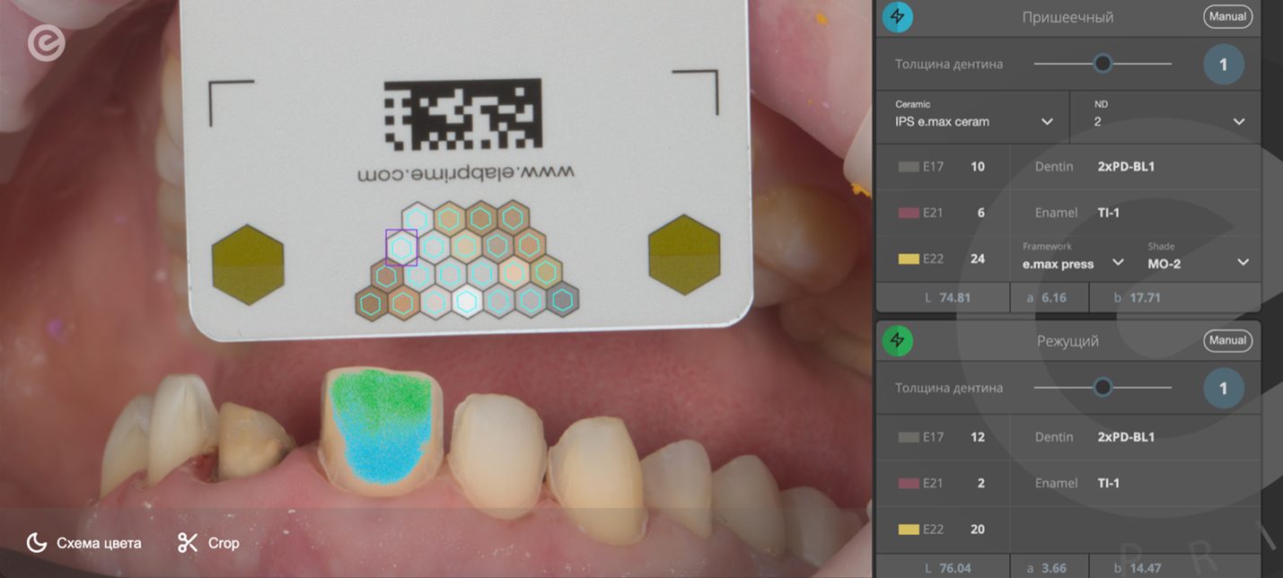
B – Definition of a porcelain recipe in the eLAB Prime software.
After shade selection, a coping was fabricated from an IPS E.max Press (Ivoclar Vivadent) ingot in shade MO-2. Following trial fitting of the coping on a master cast (Figure 5) and approval by the clinician, ceramic veneers were fabricated using Noritake EX-3 (Kuraray Noritake Dental Inc., Japan), with uniform thickness and structure (Figure 6). After verifying the fit of the veneers on the master model, the case was sent to the clinic for clinical try-in and final cementation.

Figure 5. Porcelain veneers and 21 telescope.

Figure 6. Porcelain veneers on the master cast.
After intraoral try-in of the veneers and selection of the appropriate luting shade using Variolink Try-In pastes, rubber dam isolation was applied to the operative field (Figure 7). The lithium disilicate coping was first cemented using Variolink Esthetic DC Refill (Ivoclar Vivadent AG) in the “Warm” shade (Figure 8). Both the tooth and the coping were prepared according to the adhesive cementation protocol for lithium disilicate restorations. A radiographic check was performed following cementation.

Figure 7: Dental isolation with the rubberdam for adhesive fixation.
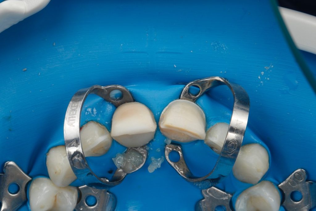
Figure 8. Fixed telescope on the tooth 21.
Subsequently, teeth 21 and 11 were restored. The lithium disilicate coping was conditioned according to the adhesive protocol for lithium disilicate ceramics, while the veneers were prepared in accordance with the adhesive bonding protocol for feldspathic ceramics. The veneer on tooth 11 was luted using Variolink Esthetic LC cement in the Light Plus shade, and the veneer on the coping for tooth 21 was cemented using Variolink Esthetic LC in the Light shade (Figure 9). For the lateral incisors and canines of the maxilla, Variolink Esthetic LC in the Neutral shade was used. The materials employed are presented in Figure 10.
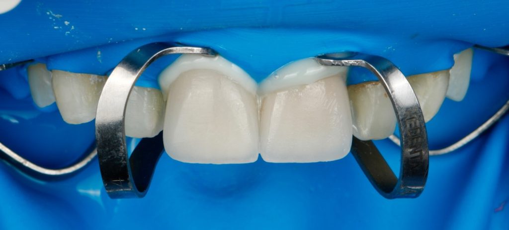
Figure 9. Porcelain Veneer 11 and 21 fixation.

Figure 10. Materials used for fixation of ceramic restorations in the presented clinical case.
Three days after tooth rehydration, the patient was recalled for a follow-up visit. Soft tissue healing following the cementation procedure was observed. An acceptable color match and integration of the ceramic veneers was also noted (Figures 11 and 12). The radiographic image of tooth 21 is presented in Figure 13.

Figure 11. Clinical view of the anterior maxillary teeth 3 days after fixation.

Figure 12. Clinical view of the smile 3 days after fixation.

Figure 13. Periapical radiograph of tooth 21 after fixation
Discussion
Despite current technological advancements in addressing tooth and restoration discoloration, the selection of a natural shade for anterior teeth remains a complex task, often resulting in uncertain outcomes. Until recently, the predictability of esthetic results relied heavily on the skill of the clinician and the expertise of the prosthodontist. The use of polarized photography and the eLAB prime shade mapping system provides the dental technician with a precise and optimized workflow for achieving reliable shade matching.
The proposed protocol is an adapted and simplified version of the method introduced by Meng et al., which combines standardized white balance and exposure correction with the latest advancements in artificial intelligence and final shade adjustments using luting cement.
Conclusion
This clinical case demonstrated that standardized digital photography utilizing the eLAB system serves as a simple and accessible tool to achieve predictable esthetic outcomes through the use of feldspathic ceramic veneers and a lithium disilicate coping.
References.
- Priyanka SR Veronica. Tooth discolouration due to endodontic materials and procedures. IOSR Journal of Dental and Medical Sciences. 2013;9:32–6
- Tsanaktsidis D. Tooth bleaching guided by elab protocol. Endodontics Today. 2021;19(4):326-329. https://doi.org/10.36377/1683-2981-2021-19-4-326-329
- Nogueira AD, Della Bona A. The effect of a coupling medium on color and translucency of cad cam ceramics. J Dent. 2013;3:18-23.
- Paravina RD, Ghinea R, Herrera LJ, et al. Color difference thresholds in dentistry. J Esthet Restor Dent. 2015;27:1-9.
- Perroni AP, Bergoli CD, Bertolini MFS, et al. Spectrophotometric anal- ysis of clinical factors related to the color of ceramic restorations: a pilot study. J Prosthet Dent. 2017;118:611-616.
- Bosenbecker J, Barbon FJ, de Souza Ferreira N, Morgental RD, Boscato N. Tooth discoloration caused by endodontic treatment: A cross-sectional study. J Esthet Restor Dent. 2020 Sep;32(6):569-574. doi: 10.1111/jerd.12572.
- Krastl G, Allgayer N, Lenherr P, Filippi A, Taneja P, Weiger R. Tooth discoloration induced by endodontic materials: a literature review. Dent Traumatol. 2013 Feb;29(1):2-7. doi: 10.1111/j.1600-9657.2012.01141.x. Epub 2012 Apr 19. PMID: 22513082.
- Attin T, Paque F, Ajam F, Lennon AM. Review of the current status of tooth whitening with the walking bleach technique. International Endodontic Journal. 2003;36:313–29.
- Pallarés-Serrano A, Pallarés-Serrano S, Pallarés-Serrano A, Pallarés-Sabater A. Assessment of Oxygen Expansion during Internal Bleaching with Enamel and Dentin: A Comparative In Vitro Study. Dent J (Basel). 2021 Aug 24;9(9):98. doi: 10.3390/dj9090098.
- Carrasco L.D., Fröner I.C., Corona S.A.M., Pécora J.D. Effect of internal bleaching agents on dentinal permeability of non-vital teeth: Quantitative assessment. Dent. Traumatol. 2003;19:85–89. doi: 10.1034/j.1600-9657.2003.00112.x
- Klarick E., Rakic M., Sever I., Milat O., Par M., Tarle Z. Enamel and dentin microhardness and chemilcal composition after experimental light-activated bleaching. Oper. Dent. 2015;40:e132–e141. doi: 10.2341/14-148-L.
- Xing W., Chen X., Ren D., Zhan K., Wang Y. The effect of ceramic thickness and resin cement shades on the color matching of ceramic veneers in discolored teeth. Odontology. 2017;105(4):460–466. doi: 10.1007/s10266-016-0287-9
- Montero J., Gómez‐Polo C. Effect of ceramic thickness and cement shade on the final shade after bonding using the 3D master system: a laboratory study. Clinical and Experimental Dental Research. 2016;2(1):57–64. doi: 10.1002/cre2.22.
- Begum Z., Chheda P., Shruthi C. S., Sonika R. Effect of ceramic thickness and luting agent shade on the color masking ability of laminate veneers. The Journal of Indian Prosthodontic Society. 2014;14(S1):46–50. doi: 10.1007/s13191-014-0362-2
- Coachman C., Gurel G., Calamita M., Morimoto S., Paolucci B., Sesma N. The influence of tooth color on preparation design for laminate veneers from a minimally invasive perspective: case report. The International Journal of Periodontics & Restorative Dentistry. 2014;34(4):453–459. doi: 10.11607/prd.1900
- Omar H., Atta O., El-Mowafy O., Khan S. A. Effect of CAD-CAM porcelain veneers thickness on their cemented color. Journal of Dentistry. 2010;38:e95–e99. doi: 10.1016/j.jdent.2010.05.006.
- Faus-Matoses V., Faus-Matoses I., Ruiz-Bell E., Faus-Llacer V. J. Severe tetracycline dental discoloration: restoration with conventional feldspathic ceramic veneers. A clinical report. Journal of Clinical and Experimental Dentistry. 2017;9(11):e1379–e1382. doi: 10.4317/jced.54359
- Greta D.C., Colosi H.A., Gasparik C., Dudea D. Color comparison between non-vital and vital teeth. J. Adv. Prosthodont. 2018;10:218–226. doi: 10.4047/jap.2018.10.3.218.
- Hein S, Modrić D, Westland S, Tomeček M. Objective shade matching, communication, and reproduction by combining dental photography and numeric shade quantification. J Esthet Restor Dent. 2021 Jan;33(1):107-117. doi: 10.1111/jerd.12641.
- Mourouzis P, Koulaouzidou E, Palaghias G, Helvatjoglu-Antoniades M. Color match of luting composites and try-in pastes: the impact on the final color of CAD/CAM lithium disilicate restorations. Int J Esthet Dent. 2018;13(1):98-109.
- Schlichting LH, Resende TH, Reis KR, Raybolt Dos Santos A, Correa IC, Magne P. Ultrathin CAD-CAM glass-ceramic and composite resin occlusal veneers for the treatment of severe dental erosion: An up to 3-year randomized clinical trial. J Prosthet Dent. 2022 Aug;128(2):158.e1-158.e12. doi: 10.1016/j.prosdent.2022.02.009.
- Hein S, Zangl M. The use of a standardized gray reference card in dental photography to correct the effects of five commonly used diffusers on the color of 40 extracted human teeth. Int J Esthet Dent. 2016 Summer;11(2):246-59.