

Abstract
The aim of the study is to carry out a comparative analysis of the phase composition of conventional zirconium with the predominance of tetragonal phase after artificial aging and loading.
The studied group included samples of conventional zirconia stabilized with 3 mol% yttrium (3Y- TZP).The X-ray diffraction analysis of these samples was carried out on an automatic X-ray diffractometer for polycrystalline materials DRON-7 in the step-by-step scanning mode. The following studies included flexural strength before and after aging, cyclic loading before and after aging, cyclic load after aging 100-200 N, 500 – 800 N, phase analysis in the area of the crack and without crack.
The diffraction patterns correlate well with each other and indicate basically the same crystal structure of the ZrO2 compound. All X-ray diffraction patterns show only the ZrO2 phase with a tetragonal crystal lattice. It was not found that the aging, cracks and loads performed affects the phase composition and microstructure of the samples under study.
The presence of cracks and loads in this case does not affect the phase composition and microstructure of the material. Samples are almost identical. It should be noted that this method is integral (volumetric) and does not allow localizing studies only in the area of the cracks and loads.
Introduction
In recent years, many ceramic materials have been introduced to the dental market. Today, the three main types of materials used to create fixed restorations include glass-ceramics (monolithic lithium disilicate; zirconium oxide framework lined with lithium disilicate, leucite and feldspathic ceramics), polycrystalline ceramics (zirconium oxide) and hybrid composites (polymer-infused ceramics, nanoceramics).
One of them is yttrium oxide stabilized polycrystalline zirconia, which has become popular in dentistry due to its adequate mechanical properties and biocompatibility.
Zirconia is mainly used as a framework for all- ceramic crowns and fixed partial dentures, usually requiring ceramic veneering and subsequent processing steps to obtain proper aesthetics because of its high opacity. Zirconia stabilized with 3 mol% Yttrium (3Y-TZP) is the first dental Y-TZP zirconia (Lüthy et al., 2005), which has exceptional mechanical properties. However, the translucency of conventional Y- TZP zirconia is no more than 70% lithium disilicate. Also, to provide enough space for the veneering ceramic to compensate for the opacity of zirconia, substantial tooth reduction is require.
In addition, the most common clinical complication of zirconia restorations is cohesive chipping of the ceramic veneer.
The final properties of zirconia will be highly dependent on both the manufacturing processing steps during fabrication of the restoration and the occurrence of post- processing surface damage due to grinding, sandblasting, and interaction with antagonists11. Also, zirconia has different surface preparation methods for fixation, different cements for fixation, and longevity problems of bonding between cements and the prepared zirconia surface.
The aim of the study is to carry out a comparative analysis of the phase composition of conventional zirconium with the predominance of tetragonal phase after artificial aging and loading.
Materials and methods
The same standardized samples (disks) of different types of zirconium dioxide with a diameter of 10 mm and a height of 1 mm were used for all experiments. Free open source software Tinkercad (Autocad) was used to create the STL file of the model. The 3D design of the model was created in the software. The base shape of the model and model parameters were selected: length, width, height, radius. Then “model holders” were added for easy separation of the milled model from the zirconium dioxide block before synthesizing. The model was exported as an STL file and then loaded into the CAD/CAM program in the dental laboratory (Exocad). The models were milled and synthesized in accordance with standard (conventional sintering). The group included samples of conventional zirconia stabilized with 3 mol% yttrium (3Y-TZP).
The X-ray diffraction analysis of these samples was carried out on an automatic X-ray diffractometer for polycrystalline materials DRON-7 in the step-by-step scanning mode. 2θ angle interval from 20° to 70° with scanning step
∆2θ = 0.02° and 3 s exposure per point. We used Cu Kα-radiation (Ni-filter), which was subsequently decomposed into Kα1 – and Kα2 – components during the processing of the spectra. The following studies included flexural strength before and after aging, cyclic loading before and after aging, cyclic load after aging 100-200 N, 500 – 800 N, phase analysis in the area of the crack and without crack.
Artificial aging (low temperature degradation) was performed using the following autoclaving regime: 134 degrees, 2 atmospheres, 5 hours.
Results
Figures 1-8 show the diffraction patterns of the studied samples.
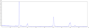
Figure 1. X-ray diffraction pattern of sample 1 (the sample was previously ground into powder).
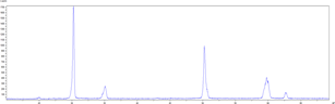
Figure 2. Diffraction pattern of sample with flexural strength before aging.
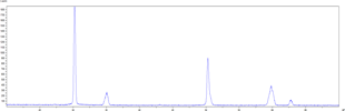
Figure 3. Diffraction pattern of sample with flexural strength after aging.
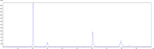
Figure 4. Cyclic loading before aging.
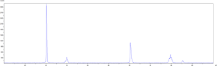
Figure 5. Cyclic load after aging 100-200 N.
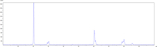
Figure 6. Cyclic load after aging 500-800 N.
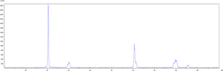
Figure 7. Phase analysis (interested in the area of the crack).
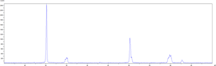
Figure 8. Phase analysis without crack.
The diffraction patterns (Fig. 1-8) correlate well with each other and indicate basically the same crystal structure of the ZrO2 compound. As can be seen from Figures 1-8, all X-ray diffraction patterns show only the ZrO2 phase with a tetragonal crystal lattice (figure 9 shows the X-ray diffraction data for this compound from the international X-ray diffraction database for polycrystalline studies IDD PDF-2.
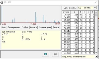
Figure 9. X-ray diffraction data of ZrO2 with tetragonal syngony (No. 17-923 IDD PDF-2).
However, in sample before heat treatment, a small amount of ZrO2 impurity with a monoclinic lattice was found (natural mineral baddeleyite, data No. 37-1484 IDD PDF-2). The diffraction pattern contains small peaks in the range of angles 2θ – 28.25o and 31.38o, which are characteristic and most intense for ZrO2 with a monoclinic lattice. After sintering, the monoclinic phase of ZrO2 completely transforms into a tetragonal phase, as can be seen from the diffraction patterns of samples 2–8, these peaks are absent. The X-ray diffraction data of IDD PDF-2 for ZrO2 with a monoclinic lattice are shown in Figure 10.
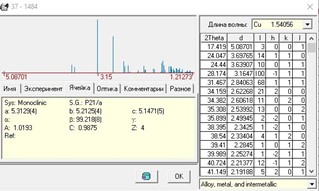
Figure 10. X-ray diffraction data of ZrO2 with monoclinic system (No. 37-1484 IDD PDF-2)
Comparing the diffraction patterns of No. 2-3 and No. 4-5, it was found that they are almost identical. Thus, within the framework of this method, it was not found that the aging performed affects the phase composition and microstructure of the samples under study. Figures 4 and 5 are almost identical.
Unlike aging, cyclic loading causes deformations in the crystal structure of the sample. The degree of deformation is directly proportional to the load. Under a cyclic load of 500–800 N (figure 6), distortions of the ZrO2 tetragonal crystal lattice in different blocks are most noticeable. Figure 12 shows, for example, the combined diffraction patterns of 5 and 6 figures (full and its enlarged fragments).
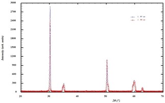
Figure 11. Combined diffraction patterns of samples with and without crack.
As the load increases, individual peaks more and more clearly split into a series of peaks, indicating deformation of the crystal tetragonal lattice of ZrO2 in different blocks and a possible decrease in symmetry. At the same time, in general, the microstructure of the material becomes somewhat better – the peaks become narrower and better resolved.
An analysis of the diffraction patterns of samples 7 and 8 on the possible effect of cracks on the phase composition and microstructure of the material within the framework of this method did not reveal any differences. The diffraction patterns are almost identical (Fig. 11).
Discussion
To quantify the phase content of zirconium dioxide samples, X-ray diffraction analysis (X-ray diffraction – XRD) is used. In X- ray diffraction analysis, the material is irradiated with incident X-rays, and then the intensity and scattering angles of the X-rays that scatter from the material13 are measured. Crystals are regular arrays of atoms, while X-rays can be thought of as waves of electromagnetic radiation. Atoms of crystals scatter incident X-rays, mainly due to the interaction with the electrons of the atoms.
Peak positions are determined by comparison with reference diffraction data. The intensities of the diffraction peaks are used as preferred phase orientations to compare different commercial grades of zirconia. The percent phase transformation obtained from the graph is based on the Rietveld14 refinement of X-ray diffraction peaks.
From technological and orthopedic points of view, phase transformation was considered to be of paramount advantage because it allows a kind of self-healing of zirconium dioxide: it allows blocking or at least preventing the propagation of microcracks and cracks in the material15. In fact, the subsequent increase in crystal volume occurs inside the material at the crack tip, which limits the crack propagation. It should be noted that at room temperature, this transformation is irreversible and localized, centered in the zone of force application (i.e., in the occlusal load region, in the zone of increased impact and). Once limited crack propagation has occurred, zirconia can no longer confine cracks in its monoclinic configuration16. In contrast, when monoclinic zirconium dioxide is reheated to 900-1000 °C (for a limited time according to manufacturers’ instructions), the phase transformation becomes reversible: through a process called “regeneration” or “annealing”, the monoclinic crystals can be moved back to the tetragonal phase, causing relaxation of compressive stresses in the material. However, after annealing, the strength of zirconia tends to decrease, and as for the optical properties, chromatic oversaturation can occur. Consequently, heat treatment at high temperature should be used with caution and only after potentially aggressive mechanical procedures (i.e., appropriate occlusal grinding, polishing, etc.)17,18.
Conclusions
The presence of cracks and loads in this case does not affect the phase composition and microstructure of the material. Samples are almost identical. It should be noted that this method is integral (volumetric) and does not allow localizing studies only in the area of the cracks and loads.
References
- Ruse ND, Sadoun MJ. Resin-composite block for dental CAD/CAM applications. J Dent Res. 2014:93(Spec No):1232– 1234.
- Lambert H, Durand JC, Jacquot B, Fages M. Dental biomaterials for chairside CAD/CAM: State of the art. J Adv Prosthodont. 2017;9: 486–495.
- Z.Khabadze, O.Mordanov, G. Davreshyan, A. Adzhieva, O. Magomedov, S. Solimanov, S. Nazhmudinov. Physical and Chemical Conditions for the Long-Term Functioning of Restorations with a Zirconia Framework. J Int Dent Med Res. 2020; 13(1): 359-363
- Lüthy, H., Filser, F., Loeffel, O., Schumacher, M., Gauckler, L.J., Hammerle, C.H., 2005. Strength and reliability of four-unit all-ceramic posterior bridges. Dent. Mater. 21, 930–937.
- Baldissara P, Llukacej A, Ciocca L, et al. Translucency of zirconia copings made with different CAD/CAM systems. J Prosthet Dent. 2010;104:6-12
- Zhang Y. Making yttria-stabilized tetragonal zirconia translucent. Dent Mater 2014;30:1195-203.
- Al-Amleh B, Lyons K, Swain M. Clinical trials in zirconia: a systematic review. J Oral Rehabil. 2010;37:641-52.
- Monaco C, Caldari M, Scotti R. Clinical evaluation of 1,132 zirconia-based single crowns: a retrospective cohort study from the AIOP clinical research group. Int J Prosthodont. 2013;26:435-42.
- Vagkopoulou T, Koutayas SO, Koidis P, Strub JR. Zirconia in dentistry. Part 1. Discovering the nature of an upcoming bioceramic. Eur J Esthet Dent. 2009;4:130-51.
- Pihlaja J, Näpänkangas R, Raustia A. Outcome of zirconia partial fixed dental prostheses made by predoctoral dental students: a clinical retrospective study after 3 to 7 years of clinical service. J Prosthet Dent. 2016;116:40-6.
- Lohbauer U, Amberger G, Quinn GD, Scherrer SS (2010) Fracto- graphic analysis of a dental zirconia framework: a case study on design issues. J Mech Behav Biomed 3(8):623–629.
- Lebedenko IYu, Dyakonenko EE, Deev MS, Sakhabieva DA, Axelrod IB. Adhesion of dental cements to zirconia restorations. Part 2. Stomatologiya. 2021;100(4):132-136.
- Mangla O, Roy S. Monoclinic Zirconium Oxide Nanostructures Having Tunable Band Gap Synthesized under Extremely Non- Equilibrium Plasma Conditions. Proceedings. 2019; 3(1):10.
- G. Garnweitner, L. Goldenberg, O. Sakhno, M. Antonietti, M. Niederberger, and J. Stumpe, Large-scale synthesis of organophilic zirconia nanoparticles and their application in organic–inorganic nanocomposites for efficient volume holography. Small. 2017; 39(9): 1626–1632.
- Zarone F, Di Mauro MI, Ausiello P, Ruggiero G, Sorrentino R. Current status on lithium disilicate and zirconia: a narrative review. BMC Oral Health. 2019;19(1):134.
- Miyazaki T, Nakamura T, Matsumura H, Ban S, Kobayashi T. Current status of zirconia restoration. J Prosthodont Res. 2013;57(4):236–61.
- Ferrari M, Vichi A, Zarone F. Zirconia abutments and restorations: from laboratory to clinical investigations. Dent Mater. 2015;31:e63–76.
- Rodrigues CDS, Aurélio IL, Kaizer MDR, Zhang Y, May LG. Do thermal treatments affect the mechanical behavior of porcelain- veneered zirconia? A systematic review and meta-analysis. Dent Mater. 2019;35(3):807–17.