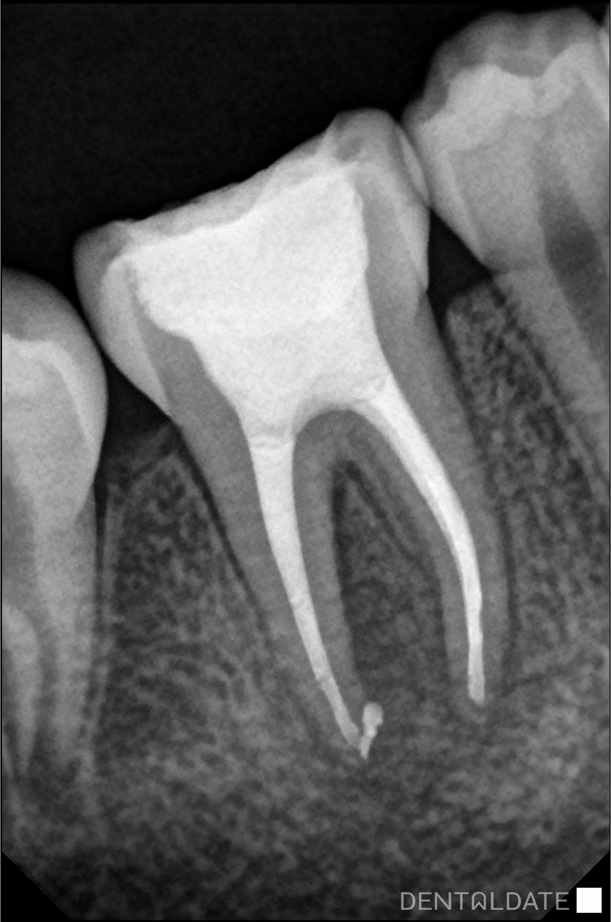
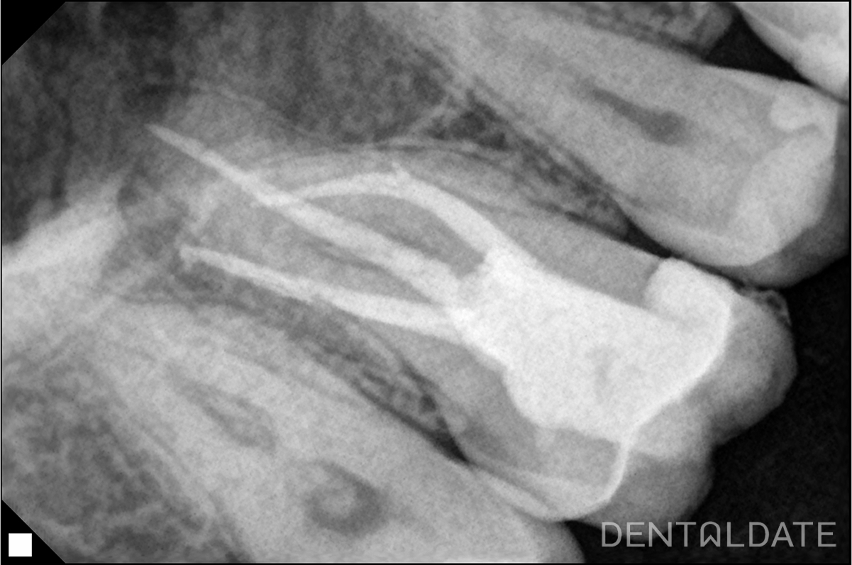
A CBCT scan was performed, revealing an area of inflammation (granuloma) near the root of tooth 1.5. The tooth had a large, failing filling.
In the upper third of the root, there was a fiber post, and the canal had been inadequately treated: it was not filled up to the apex, with voids and porosities in the root canal filling material. As a result, inflammation developed in the periapical tissue.
Treatment process:
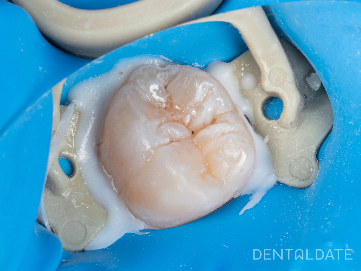
- The failing filling and underlying decay were removed.
- The lost walls of the tooth were restored using composite material (build-up).
- The fiber post was extracted, and the remnants of the old root canal filling were removed.
- The canal was mechanically and chemically cleaned.
- Calcium hydroxide medication was applied for two weeks to disinfect the canal.
- The canal was then prepared and sealed using the warm gutta-percha condensation technique.
The entire treatment was performed under a microscope for maximum precision.
Due to the initial structural damage of the tooth, it was additionally reinforced with a post and later covered with a crown for long-term strength and protection.
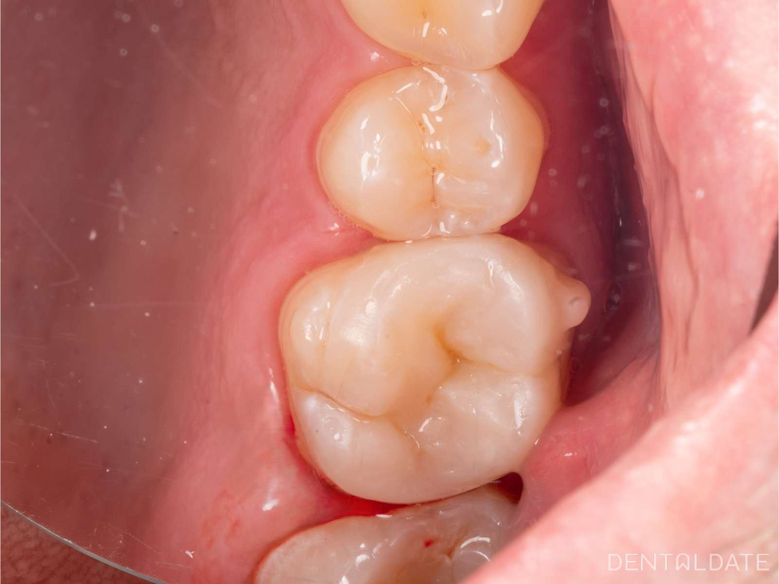
Before

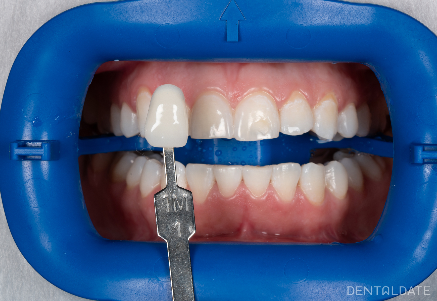
After