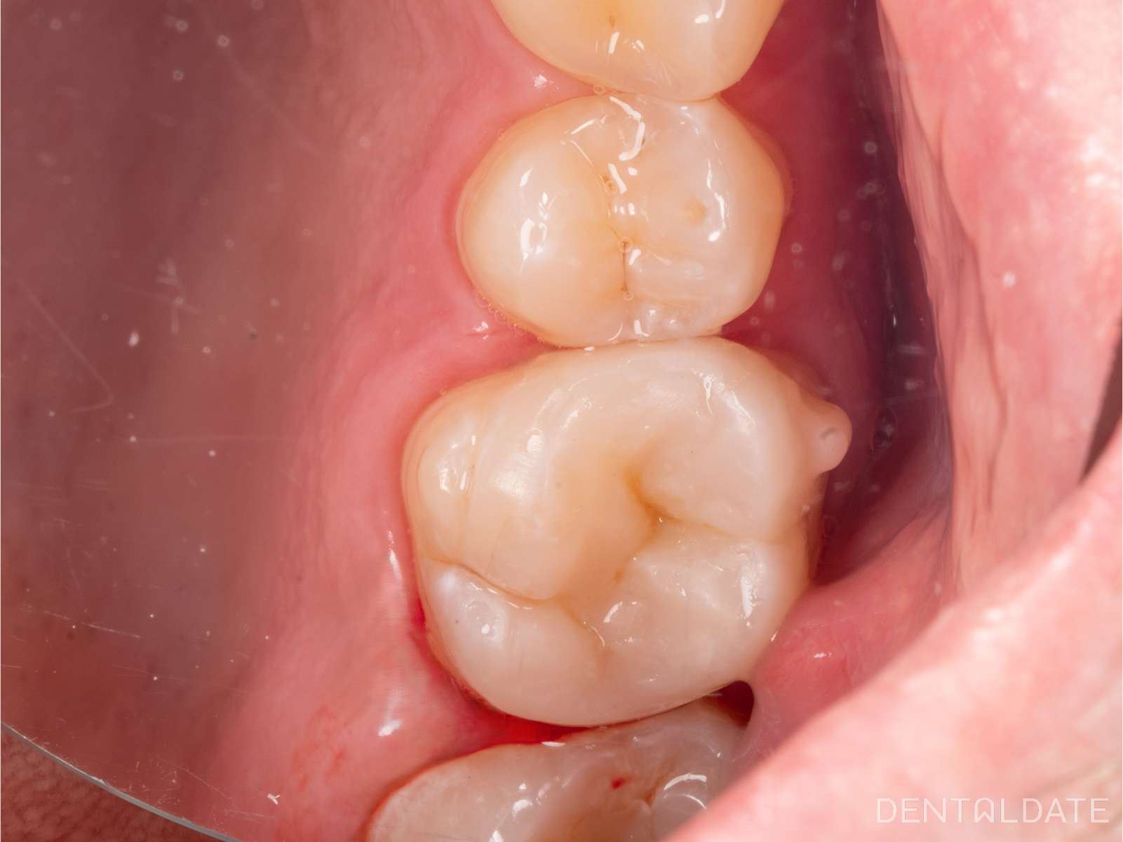
Clinical examination:
- The tooth had an intact filling on the occlusal surface.
- There was a sharp pain reaction to cold stimuli.
- A periapical X-ray revealed that the filling was placed close to the pulp horn.
Treatment process:
- Using a microscope, a precise access opening was created to the pulp chamber, preserving as much healthy tooth structure as possible.
- The infected pulp (nerve) was removed.
- The root canals were mechanically and chemically prepared and sealed in a single visit using the warm gutta-percha condensation technique.
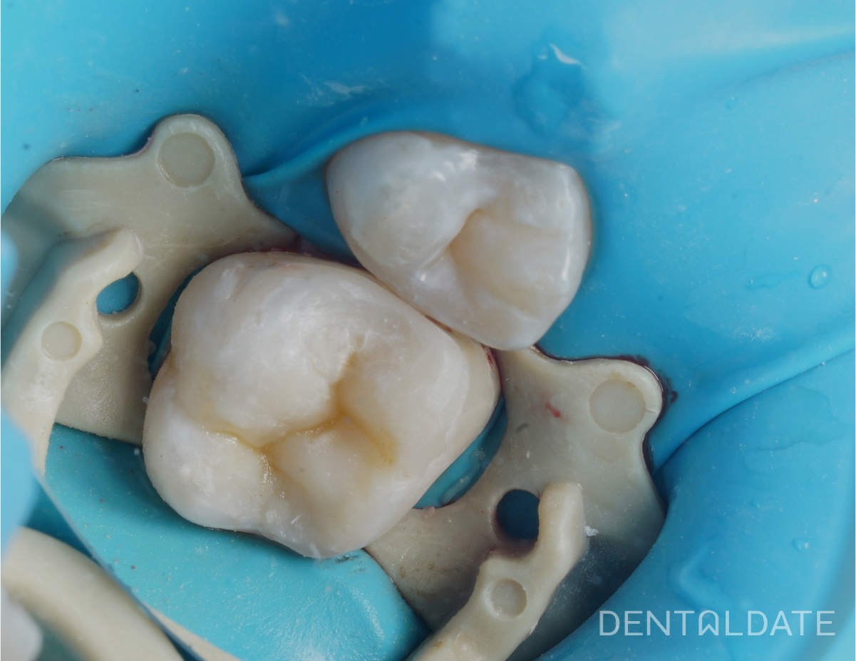
- The access cavity was restored with a composite filling.
The patient left the clinic pain-free and with a fully treated tooth.
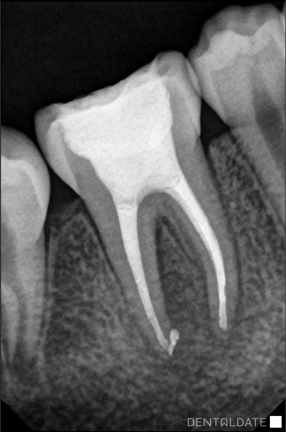
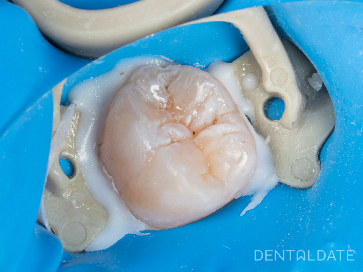
Tooth before endodontic treatment. The filling is close to the pulp horn.

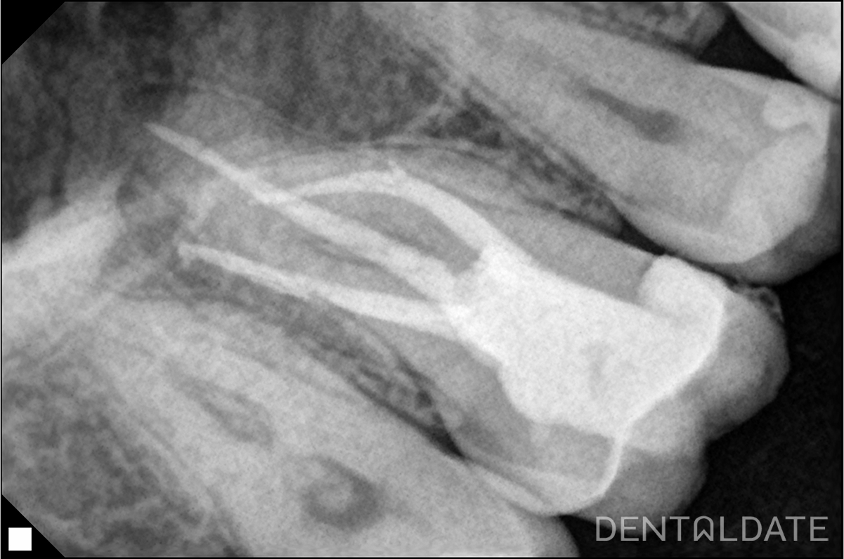
Tooth after treatment with obturated root canals.