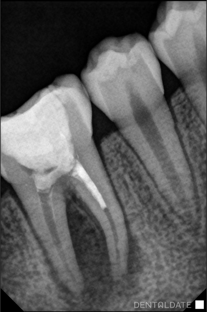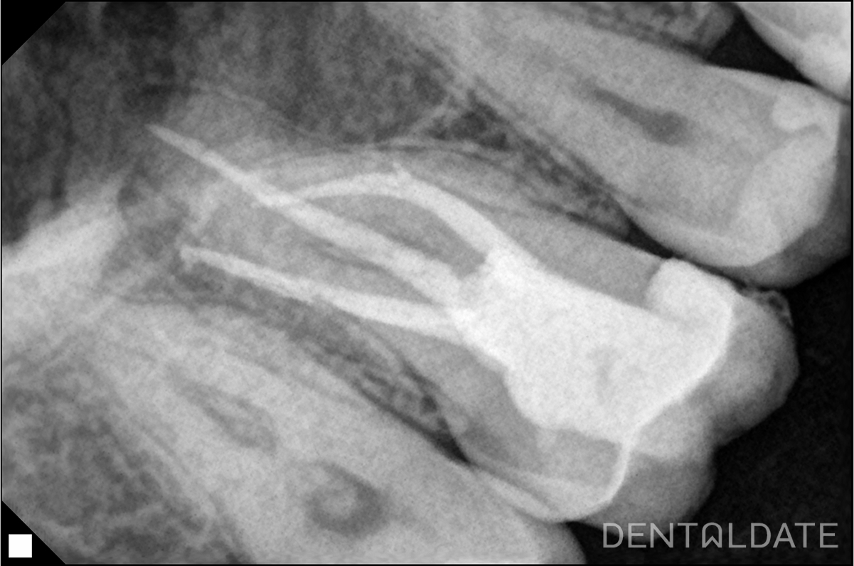

A computer tomography was performed. On which it was revealed that the tooth was previously treated endodontically. According to the patient’s mother, about a year ago due to a complication of caries (pulpitis).
The canals are under-filled by 1/2 of the root length. The material in the canals is traced intermittently, pores inside. In the periapical tissues of both roots there is a focus of bone tissue loosening. Round shape, the contours are indistinct.
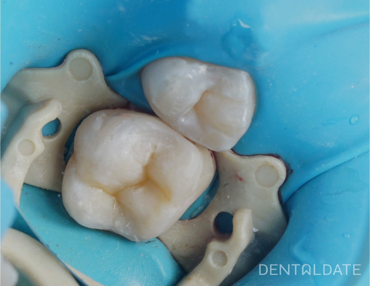
On examination, a fistulous passage was found in the projection of the roots of tooth 4.6 from the vestibular side. Tooth 4.6 under a volumetric filling, marginal adherence of the filling is disturbed. Percussion is weakly positive.
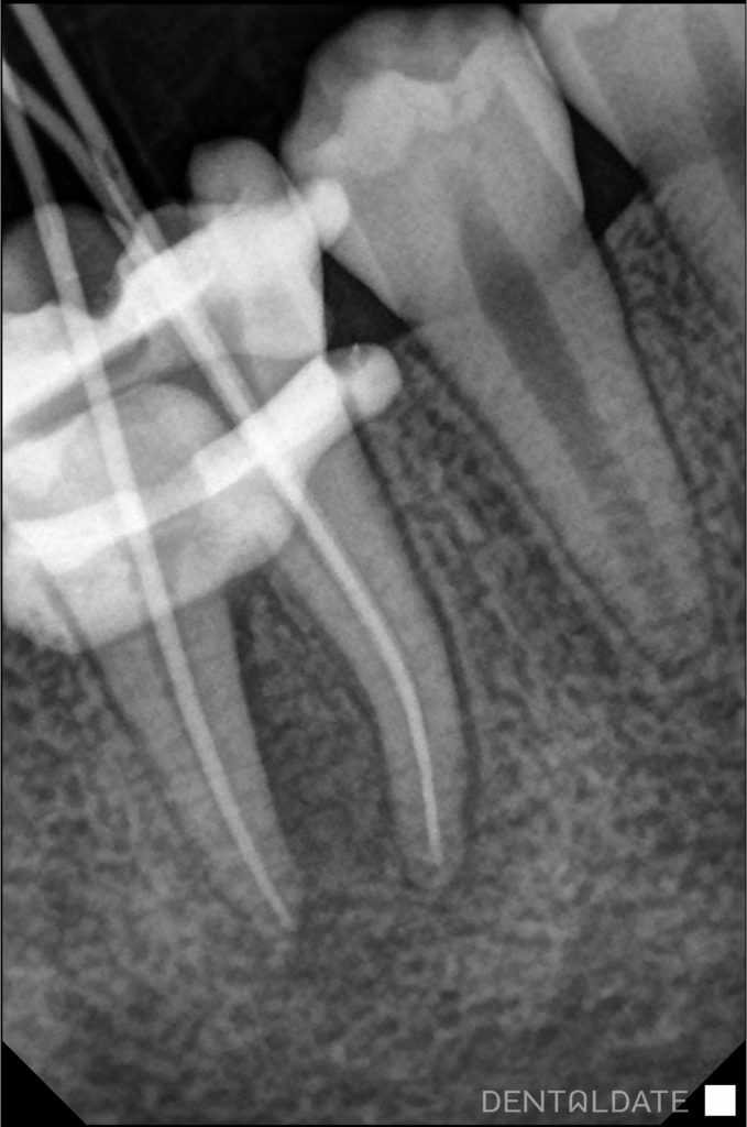
Endodontic re-treatment of tooth 4.6 was performed. The root canals of the tooth were filled, passed and treated for the whole length of the root, with intermediate placement of Calcium hydroxide in the canals for 3 weeks.
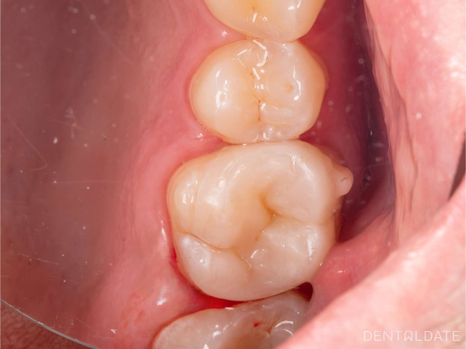
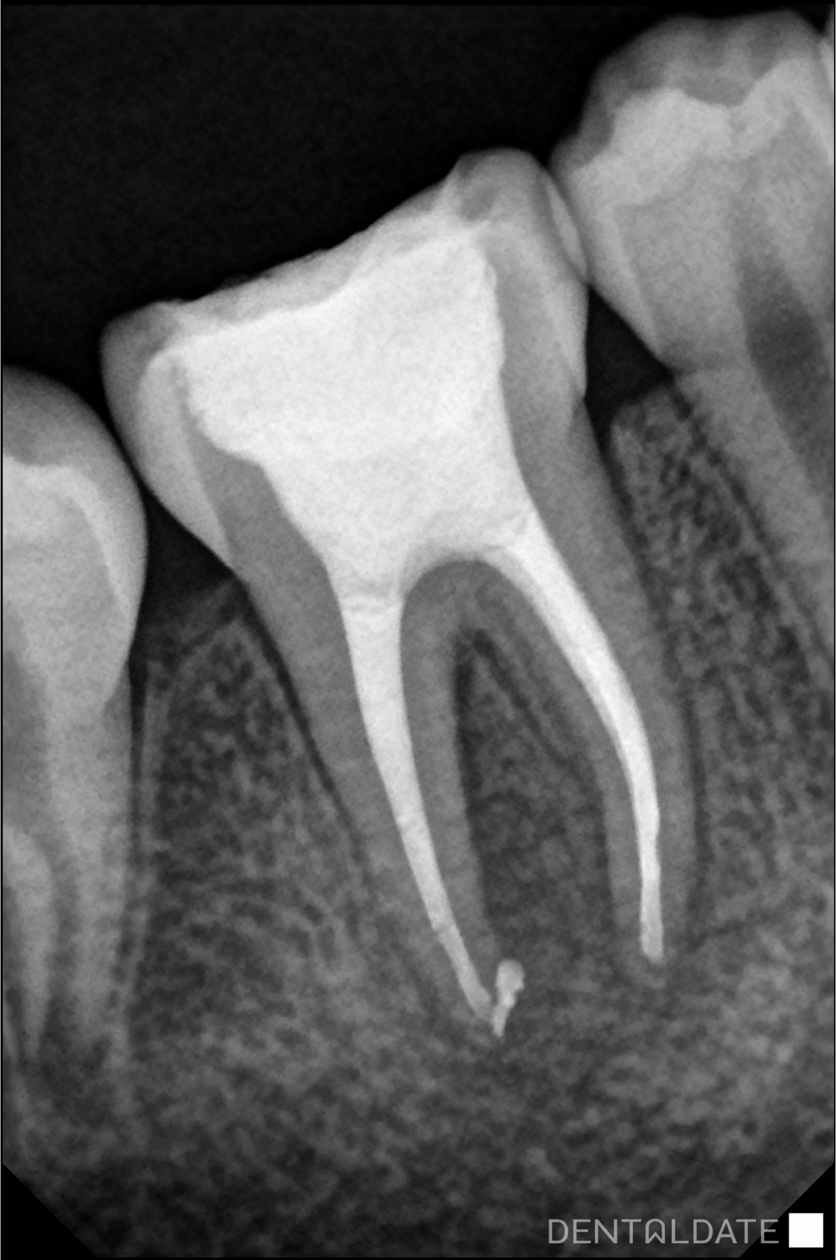
After a period of time the fistulous passage disappeared. A periapical photograph was taken, which showed a marked improvement of the rarefaction in the bone around the roots of the tooth.
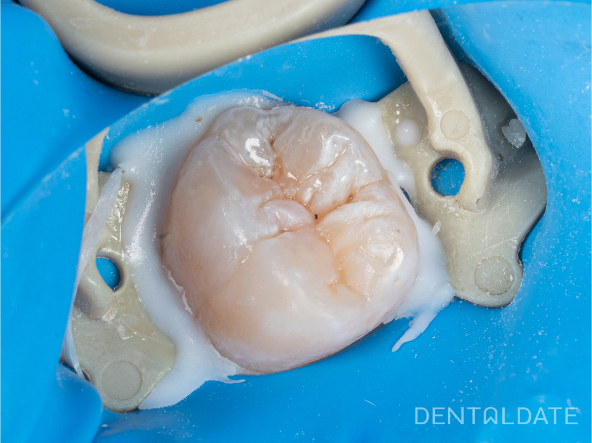
The root canals were finally prepared and filled with hot compaction gutta-percha and AHplus siler. A hermetic filling was placed. Appearance for a control scan after 6 months.