
Key Facts of This Case
Before going into detail, here is what this case involved:
- Tooth: lower wisdom tooth (38), in a deep and complex position.
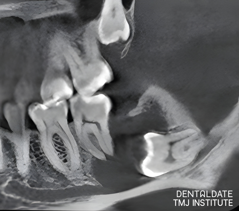
- Finding: a dentigerous (follicular) cyst of about 1 × 1 cm around the tooth.
- Main difficulty: the tooth lay right in the mandibular nerve canal.
- Treatment: removal of the tooth and the cyst together, in a single procedure of about one hour.
- Confirmation: the diagnosis was confirmed by histological examination.
- One week after surgery: the patient felt well, and the sutures were removed.
What the CBCT Showed
A regular two-dimensional X-ray can show that a wisdom tooth is impacted, but it cannot show precisely how the tooth relates to the structures around it. Cone-beam computed tomography (CBCT) gives a three-dimensional picture instead. Here it showed the lower wisdom tooth (38) lying deep, right in the mandibular nerve canal, with a cyst of about 1 × 1 cm around it. This information was essential for planning how to reach the tooth and the cyst safely.
What Is a Dentigerous Cyst?
A dentigerous, or follicular, cyst is a fluid-filled sac that forms around the crown of a tooth that has not erupted. It develops from the tissue that normally surrounds a tooth while it is still forming inside the jaw. It is a relatively rare condition and is most often associated with wisdom teeth.
Such cysts usually grow slowly and often cause no pain, so many people only learn about them from an X-ray or a scan. As a cyst enlarges, it can thin the surrounding bone and push on neighbouring teeth, which is why removal is generally recommended rather than simple observation.
Working Next to the Mandibular Nerve
The mandibular canal runs through the lower jaw and carries the nerve that gives sensation to the lower lip, the chin and the lower teeth on that side. When a wisdom tooth lies directly in this canal, the surgeon has to remove both the tooth and the cyst completely while protecting the nerve. That demands careful planning on the scan, a controlled technique and experience in complex oral surgery. It is the main reason a case like this is very different from a routine extraction.
The Operation and the Histology Report
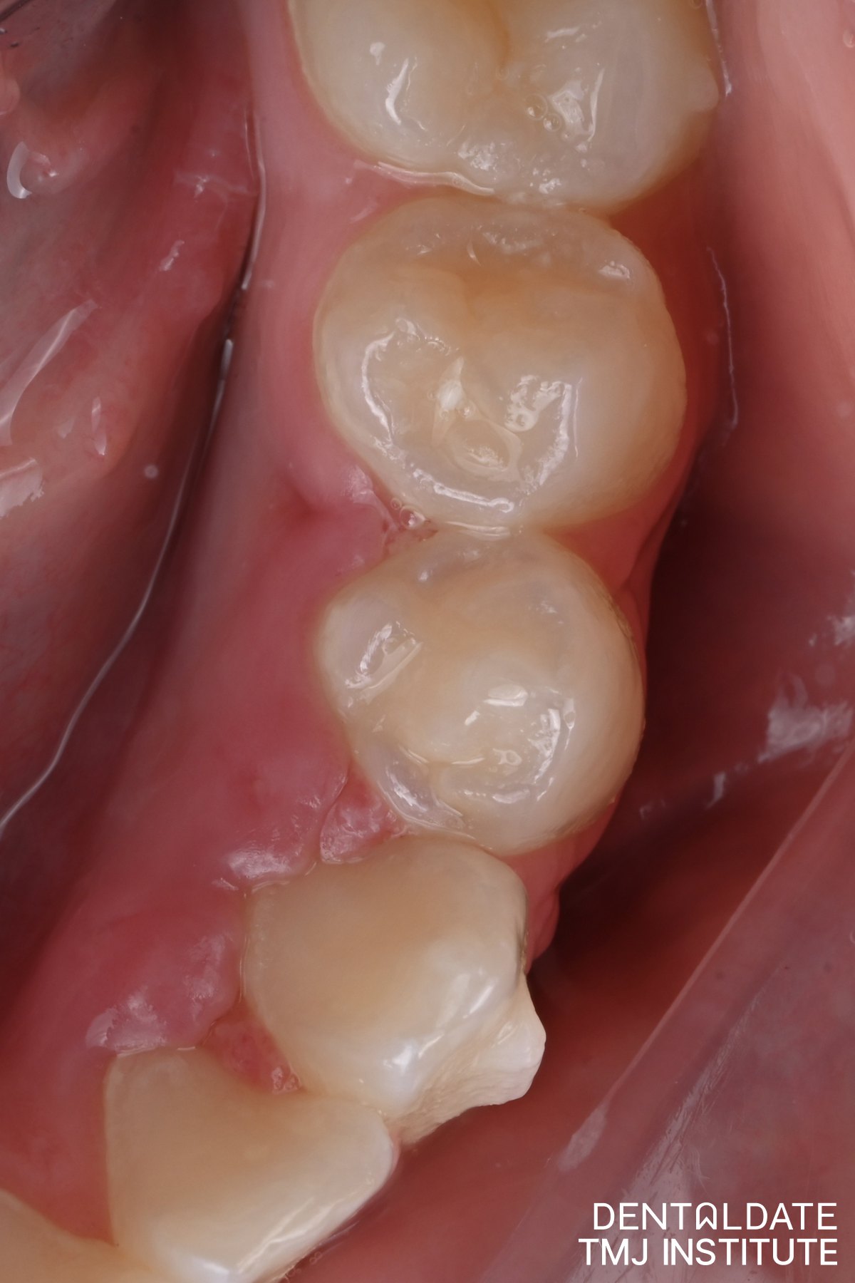
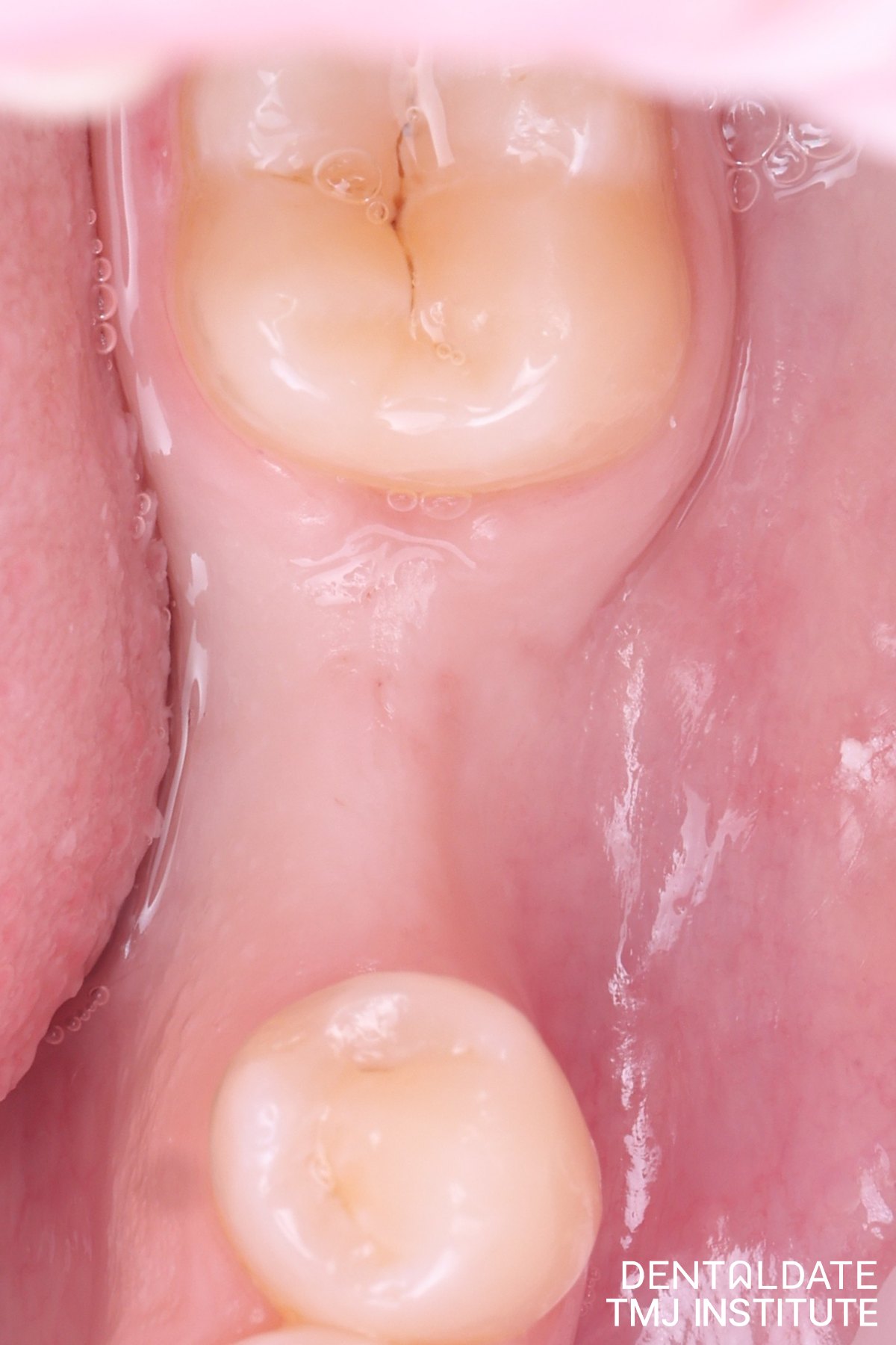
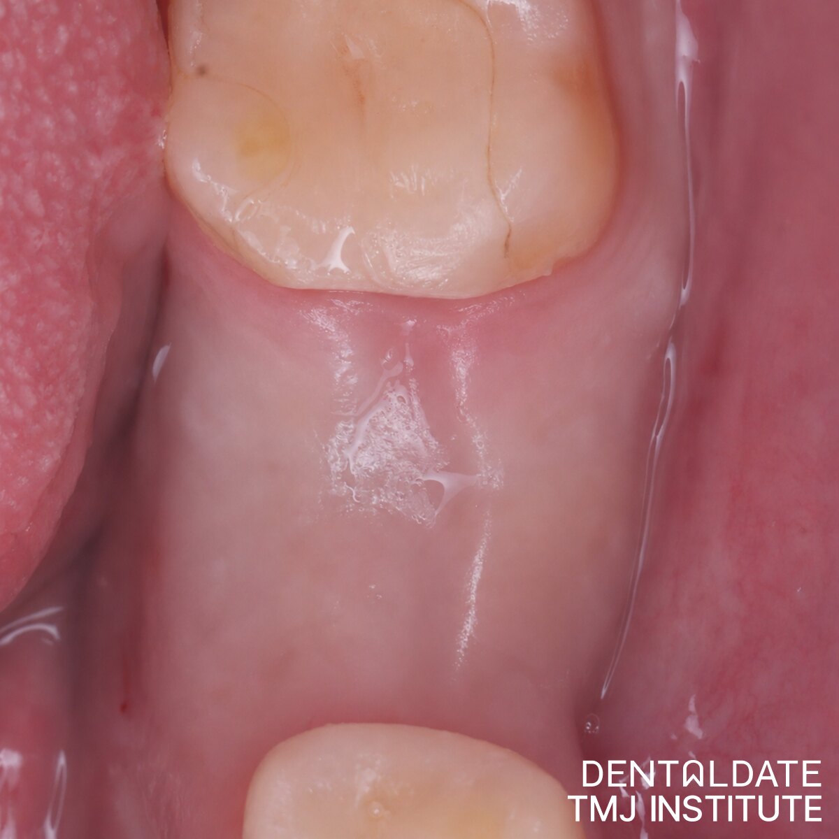
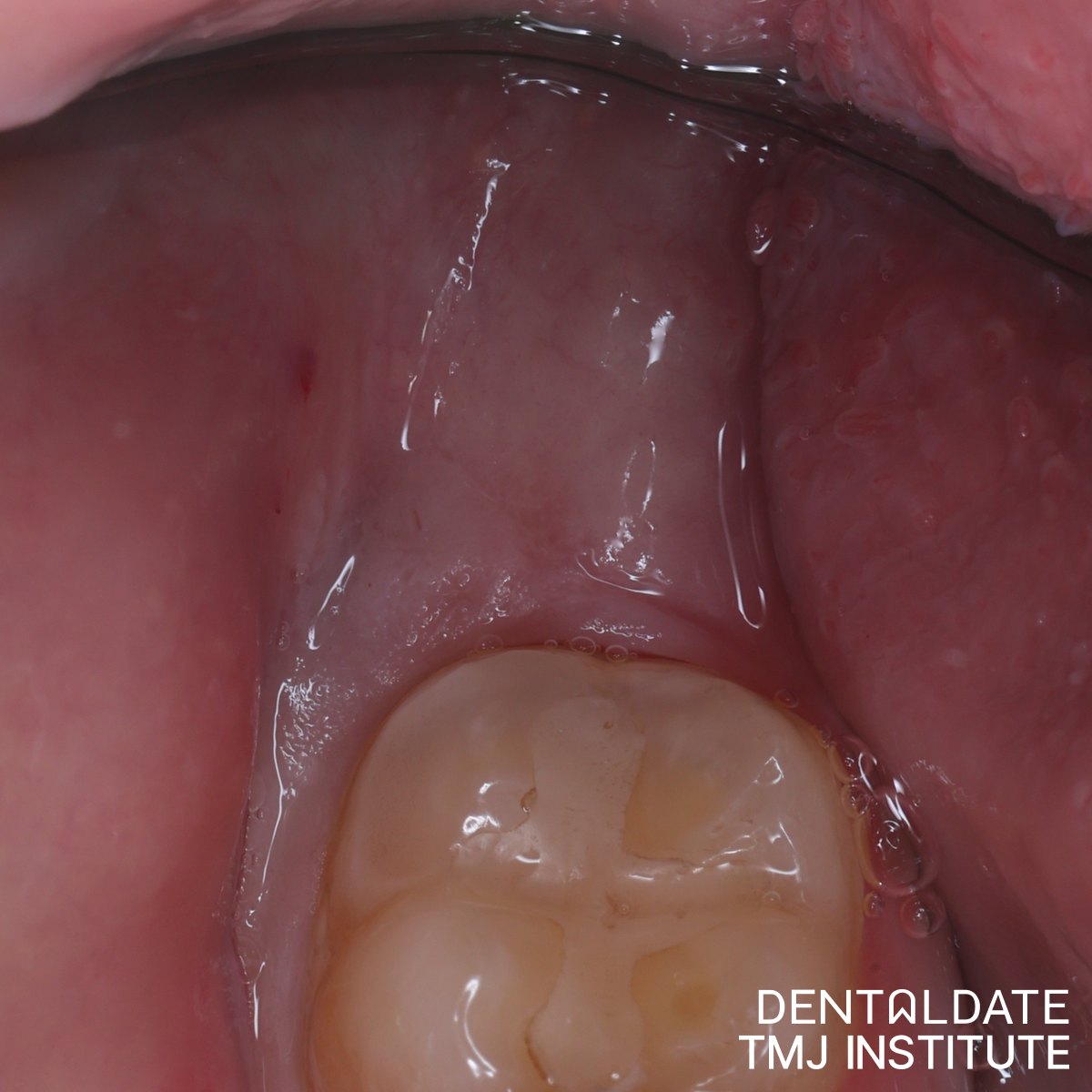
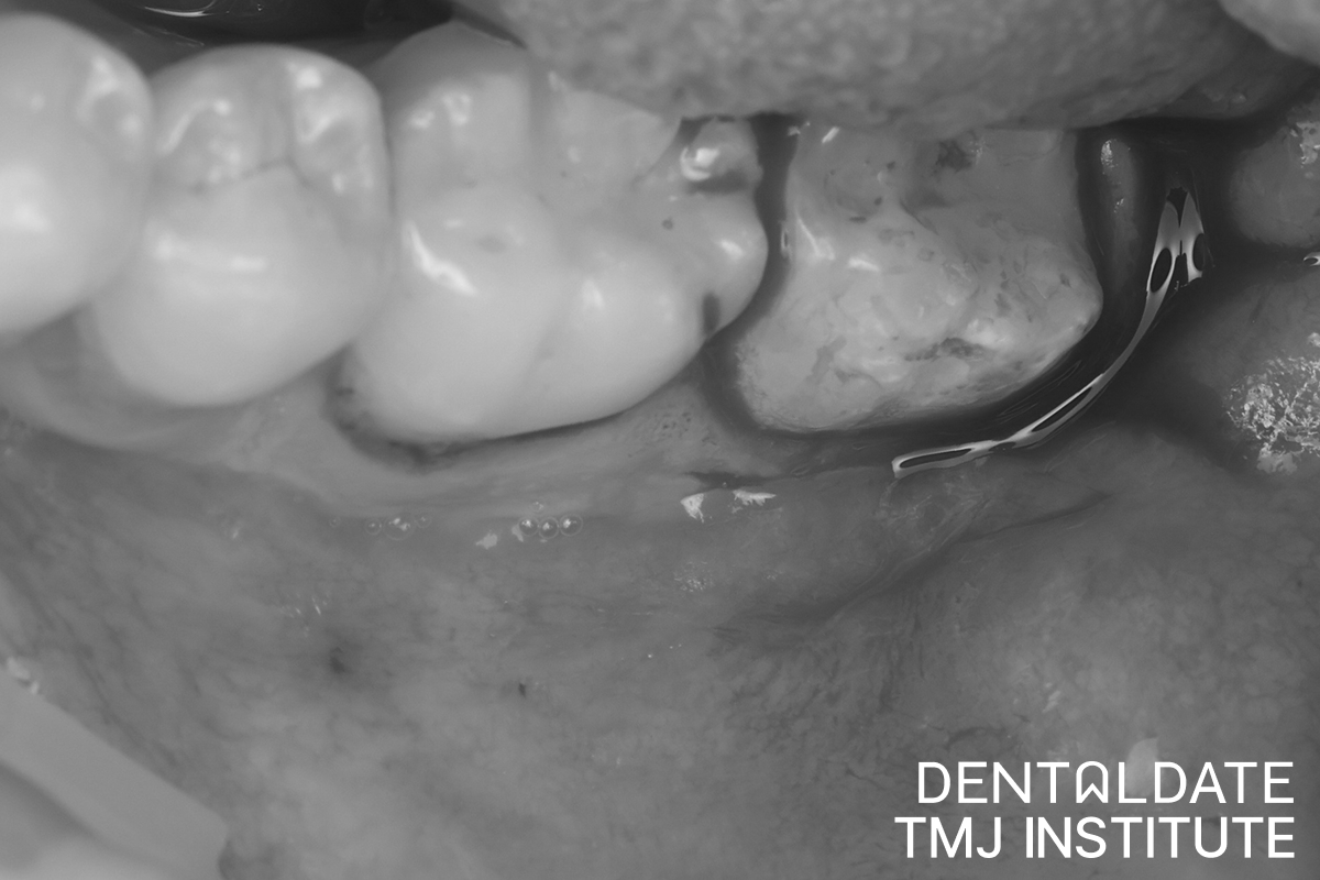
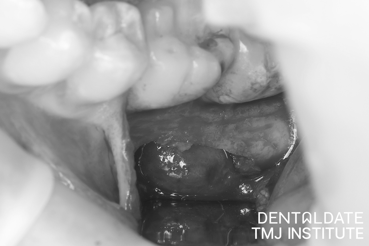
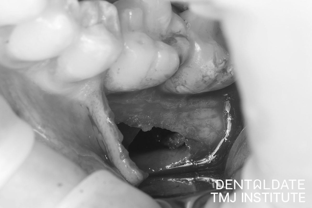
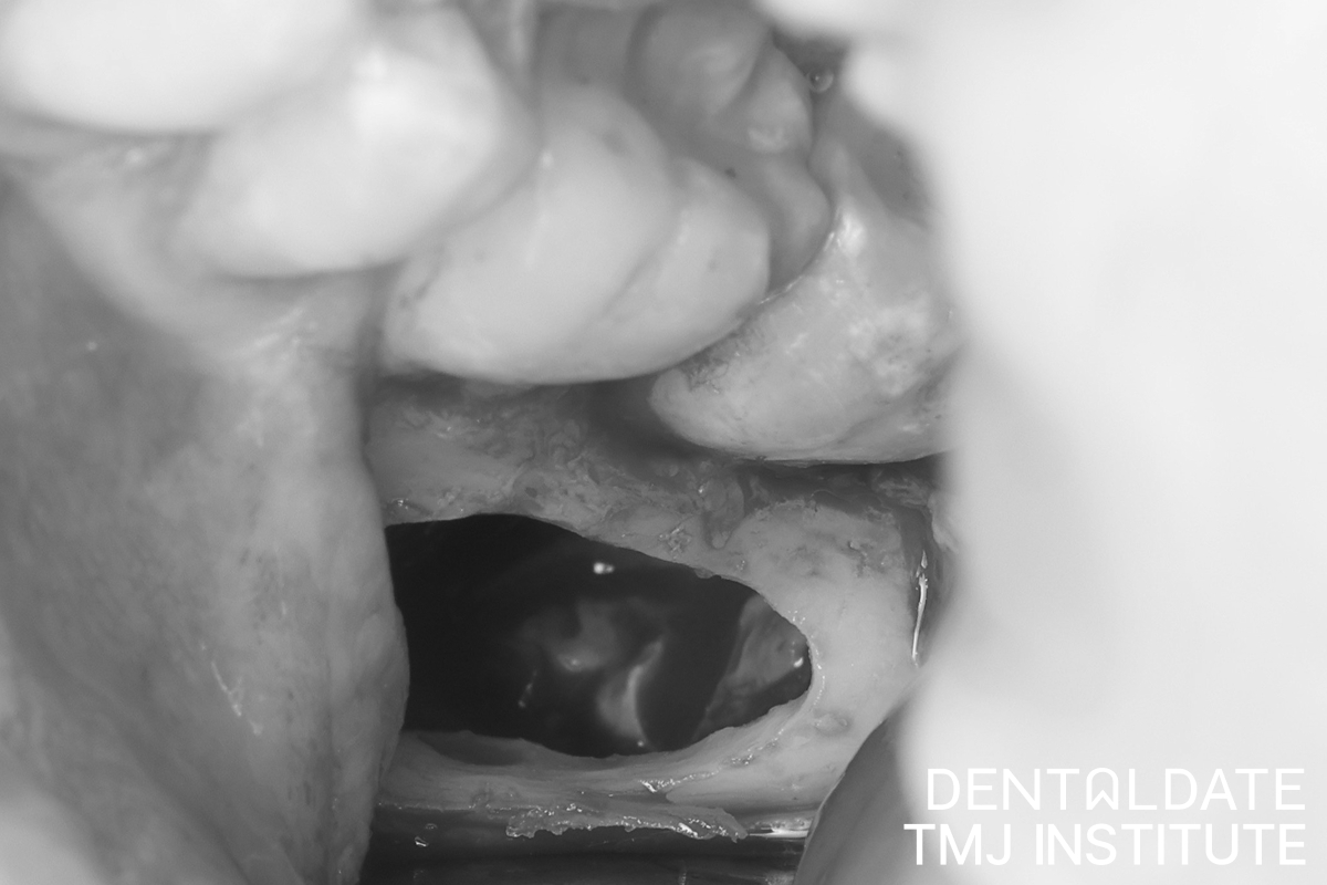
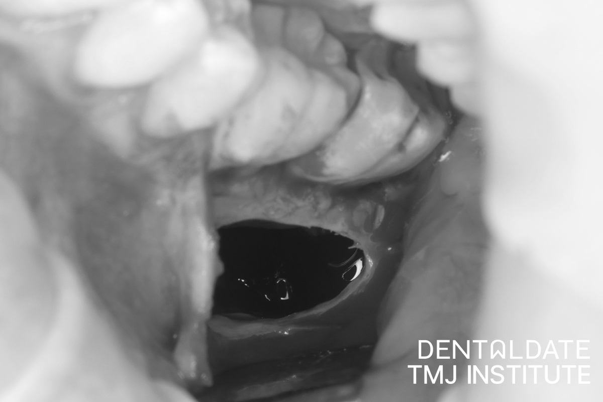
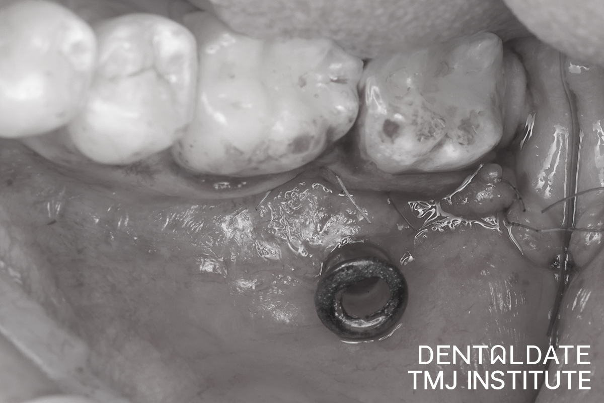
The wisdom tooth and the cyst were removed in a single procedure, which took about an hour. Removing them together meant the patient needed only one operation. At the end, the wound was closed with sutures.
The removed tissue was sent for histological examination, a laboratory study of the tissue under a microscope. This step matters because several different jaw lesions can look alike on a scan, and only histology gives a definite answer. In this case, it confirmed the diagnosis of a dentigerous cyst.
Recovery: The First Week
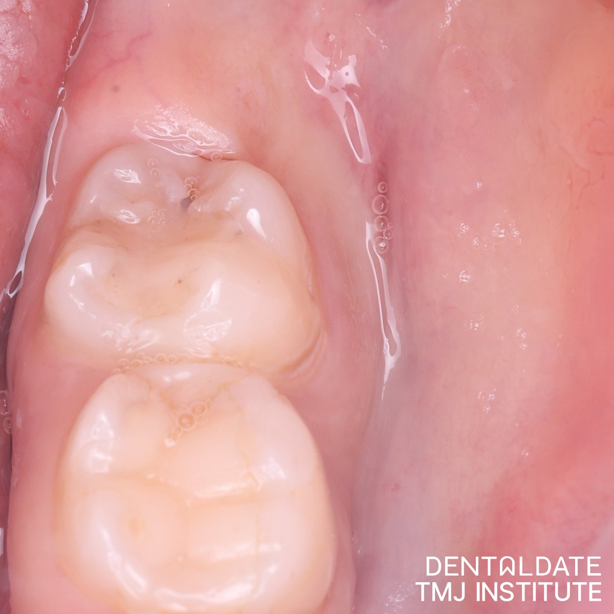
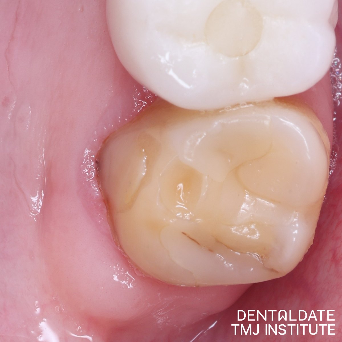
To prevent complications after removing a wisdom tooth like this, the patient took antibiotics and followed the doctor’s recommendations strictly. One week after surgery, the patient reported feeling well, and the sutures were removed.
After deep wisdom tooth surgery in general, some swelling and tenderness in the first days are a normal part of healing. Patients are usually advised to eat soft food, avoid hot drinks and strenuous activity at first, avoid smoking and keep the area clean as instructed. Unusual numbness, increasing pain or a fever should be reported to the surgeon.
Signs That Deserve an X-ray
Because a cyst around a wisdom tooth often causes no symptoms for a long time, it is usually discovered by chance. It is worth asking your dentist about imaging if you notice:
- a wisdom tooth that has never appeared in the mouth;
- a feeling of pressure or fullness at the back of the jaw;
- swelling of the jaw or of the gum near the back teeth;
- neighbouring teeth that seem to be shifting;
- numbness or tingling in the lower lip or chin.
None of these signs proves that a cyst is present, but each one is a good reason for an examination.
Planning a Difficult Extraction at DentalDate
At DentalDate, complex extractions are planned with 3D imaging, so the surgeon knows where the tooth, any cyst and the nerve are before the procedure begins. If you have been told that your wisdom tooth is in a difficult position, or a scan has shown something unusual around it, book a consultation with our oral surgery team. We will go through the images with you and explain what the next step should be.